A Box Full of Surprises
El cerevell humà i la neurogènesi
L'existència de neurogènesi adulta al cervell de mamífers, inclosa la nostra espècie, així com la identificació de les cèl·lules responsables d'aquesta neurogènesi, ha ajudat a capgirar la idea que teníem de l'organització del nostre cervell. El descobriment de l'existència de cèl·lules mare al cervell adult responsables d'aquesta neurogènesi obre noves perspectives en el camp de la medicina regenerativa, pel potencial per a restaurar àrees malmeses, però abans hem de ser capaços d'establir un diàleg precís amb aquestes cèl·lules.

|
The existence of adult neurogenesis in the brain of mammals, our species included, as well as the identification of the cells responsible for said neurogenesis, has completely changed the idea we had of our brain’s organisation. The discovery of the existence of stem cells in the adult brain responsible for said neurogenesis opens up new perspectives in the field of regenerative medicine, due to their potential to restore damaged areas, but, to get to this point, we must be able to establish a precise dialogue with them. |
||
|
Despite the great efforts of researchers to find out how our brain works, it seems that we are still taking the first steps. This century started after the so-called «Decade of the brain», a time that, when it ended, left us more questions than answers. This fact has given rise to many researchers proposing that, more than a decade, what we need is a «Century of the brain». Despite it not being defined as such, it may be this century, since in recent years there have been spectacular advances that have even torn down some of the pillars on which neuroscience was based. Perhaps one of the biggest surprises has been the discovery of the existence of neural stem cells in the adult brain of all mammals, including our own species. These cells have a precise location and nowadays we are able to extract them, cultivate them, increase their number… but we still cannot overcome one of the greatest challenges, that of establishing a precise dialogue with them. In experimental animals, these «conversations» have already been initiated. In these, through the use of some molecules or differentiation factors, we are starting to influence and guide them, so that they produce cells that are capable of joining existing damaged circuits. This is a major breakthrough, since in the future these cells could be used in therapies against neurodegenerative diseases such as Alzheimer’s, Parkinson’s or other diseases. However, we must be aware of the need for a great research effort, since this reality is still far from being achieved in the human species. For this reason, perhaps before we start controlling them, we should ask ourselves what function these stem cells have in the adult brain. |
|
|
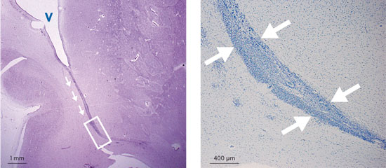
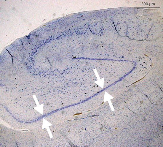 © Jose Manuel García Verdugo and Arantxa Cebrián Silla Panoramic photograph of the human hippocampus. The arrows indicate the cellular band of the dentate gyrus where the stem cells and the new neurons are found. Most of the adult neurogenesis takes place in this region. |
«This adult neurogenesis could be an adaptive mechanism that would contribute to the generation of temporal and spatial memories» |
|
|
STEM CELLS IN ADULT BRAINs Today we know that, in adults, these stem cells, far from being at rest, are active and play a role: the generation of new neurons or neurogenesis. In our species, most of the adult neurogenesis is found in the hippocampus, a region located in the medial region of the temporal lobe that is closely linked to the learning and memory processes. Within the hippocampal formation, the stem cells are located in a particular structure that is called dentate gyrus. These stem cells have been described as cells of the astroglial type (cells involved in the care and support of the central nervous system). The stem cells of the dentate gyrus are responsible for giving rise to a type of neurons called granule neurons. These new neurons are integrated into the existing hippocampal circuits and, while we know their integration mechanism, we still have many questions about what role these new neurons play in the human brain. However, studies with rodents and the application of mathematical models has enabled us to note that this adult neurogenesis could be an adaptive mechanism that would contribute to the generation of temporal and spatial memories. Which is to say that the integration of new neurons in the established circuits would facilitate the discrimination between similar temporal or spatial contexts. Therefore, this phenomenon could be favouring the adaptation to changeable situations such as seasonal changes, environmental changes, or even exposure to adverse situations such as stress. Globally, we know that the hippocampus is not an isolated structure, but it is widely interconnected with various regions, the most important being the cerebral cortex. These connections are extremely complex, since they can be both activating and inhibitory, as well as highly plastic in nature, that is, they can be modified continuously. The emergence of neurogenesis in the hippocampus has forced the expansion of this concept of plasticity and has added a new level of complexity to these connections. This way, if we imagine that the brain is a computer, the dentate gyrus, with its granule neurons, would be like a microchip in which «electronic circuits» would begin to form, functioning as the base or gateway of new information. These new circuits transmit the incoming information to other regions of the hippocampus, and these, in turn, to our brain’s «hard disk», the cortex, where information is processed and stored. Once there, the new information may connect with other previously stored information or even be stored separately, the same way we introduce new files in our computers in previously created or new folders. |
||
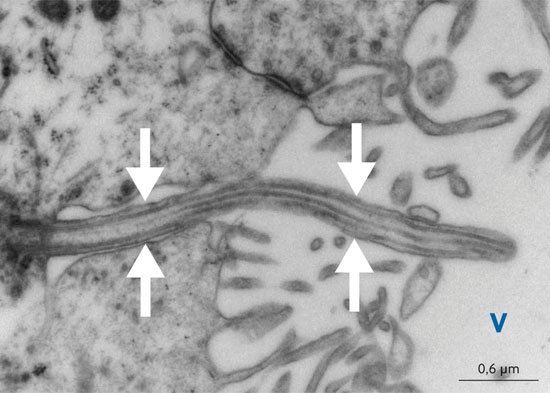 © Jose Manuel García Verdugo and Arantxa Cebrián Silla Primary cilium (marked with arrows) seen under the transmission electron microscope and corresponding to a stem cell of the lateral ventricles (V) of a rodent. This tubular structure concentrates on its surface receptors related to the proliferative cycle. The morphologic alteration or modification of this structure blocks neurogenesis. |
«Most of the adult neurogenesis is found in the hippocampus, a region located in the medial region of the temporal lobe that is closely linked to the learning and memory processes» |
|
|
Another brain region where stem cells have been identified is located in the walls of the lateral ventricles. Just as in the dentate gyrus, the nature of these cells corresponds to the astroglial type. In rodents, they produce new neurons that migrate several millimetres (at 50 µm per hour) to reach the olfactory bulb. This migration presents a characteristic organization in which cells migrate, forming chains, through the inside of cellular tubes that serve them as guide and isolate them from the rest of the nervous system. When they have reached the olfactory bulb, the migratory cells get out of these tubes, migrate radially and are integrated as immature neurons in the preestablished circuits and then change into neurons of the granule and periglomerular type. In rodents, this process takes place throughout the life of the animal, and some studies have shown that the adult neurogenesis in the olfactory bulb has an important role in the establishment of olfactory memories. Another interesting advance recently made in the study of stem cells has been in the interpretation of the function of a structure known as the primary cilium and which is characterised by a long tubular structure. This cilium concentrates on its surface receptors related to the proliferative cycle that work as a receiving antenna, and it has been shown that its morphologic alteration or modification of its receptors blocks the neurogenesis. This discovery has been applied to the field of oncology, since it has been observed that cancer stem cells also have a primary cilium. SURPRISES IN THE HUMAN BRAIN In the case of the human species, the migration to the olfactory bulb in adult stages is virtually undetectable, since, although some authors believe that there is an individualised cell migration, no cellular chains or tubes similar to those described in rodents have been found. Even though there is no migration of new cells to the human adult olfactory bulb, there is evidence that there are stem cells that persist in the walls of the lateral ventricles. In some diseases or disorders, such as amyotrophic lateral sclerosis (ALS) or cerebral infarction, stem cells present in the ventricles are activated and proliferate, producing new cells that migrate to the area of the injury. We do not know the degree of incorporation of these new cells and whether or not their role is relevant against diseases. If in non-pathological conditions our stem cells from the lateral ventricles proliferate but there is no migration to the olfactory bulb, then what is the function of this proliferation? The answer may be that most of the time we focus on the production of neurons and stem cells also produce other types of cells, such as oligodendrocytes. These are the cells responsible for forming myelin along the axons, protecting them and giving them support. This is still an underdeveloped field, and it needs long-term research. |
||
|
|
«One of the latest surprises that we found in the human brain is a massive neuronal migration that takes place in the earliest stages after birth and that fully involves the prefrontal cortex» |
|
|
Perhaps one of the latest surprises we found in the human brain is a massive neuronal migration that takes place in the earliest stages after birth and that fully involves the prefrontal cortex. During the human embryonic development, multiple migrations of new neurons towards all regions of the brain are produced, including to the olfactory bulbs. After birth and during the early breast-feeding stages (approximately up to three years of age), the majority of migrations disappear but migrations to the olfactory bulbs remain, with an identical organisation (chain migration through cellular tubes) to the one observed in rodents and non-human primates and that would be responsible for the definitive organisation of the olfactory bulbs. What is surprising is that, in the case of humans, the migration path to the olfactory bulbs forks, giving rise to a new migration corridor ending in the prefrontal cortex. This interesting migration of new neurons presents the same morphological characteristics as those previously described in the olfactory bulbs, although the shortage of this type of material has not yet allowed the study of the differences that might exist at a genetic and molecular scale. Learning about the cellular and molecular mechanisms by which these new neurons decide to separate and take an alternative route to the olfactory bulbs could be a very important step for the knowledge about the evolution of the human brain in comparison to rodents, who do not exhibit this migration to the prefrontal cortex. This is one of the most important areas of the cortex or human «hard disk». It is located in the most frontal part of the brain and is involved in many superior brain functions, such as perception, attention, memory (working memory or operative memory), language, decision making, etc. These functions, along with those of the anatomical areas of the language, characterise the human brain, differentiating it from the rest of mammals. |
||
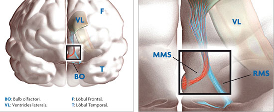 © Jose Manuel García Verdugo and Arantxa Cebrián Silla Virtual 3D drawing of the unweaned human brain, showing the migratory chains that are headed toward the olfactory bulb (RMS) and the prefrontal cortex (MMS). The cells migrate forming chains inside the cellular tubes up until they get to the olfactory bulb, where they are integrated as immature neurons in the preestablished circuits. Subsequently, these cells differentiate into granule and periglomerular neurons. In the case of the human brain, and in contrast to the brains of rodents, this migration is virtually undetectable in the adult stage. |
«The existence of adult neurogenesis in the brain of mammals, as well as the identification of the cells responsible for said neurogenesis, has completely changed the idea we had of our brain’s organisation» |
|
|
Although the field of adult neurogenesis and characterisation of stem cells to dialogue with them is growing exponentially, we must not forget the researchers who have contributed the most to the current knowledge on the role of adult neurogenesis. Firstly we must mention Joseph Altman, pioneer in describing the neurogenesis in the dentate gyrus and the migration of new neurons to the olfactory bulb in rodents, and Fernando Nottebohm, because he was the first researcher who linked neurogenesis with functionality. Their work was conducted in songbirds, where it was found that new neurons were essential for the canaries to learn new songs, which is fundamental for their reproduction. The most interesting thing is that each year they need new neurons to learn new songs and «delete» the old. Arturo Álvarez-Buylla, who, along with Joseph Altman and Giacomo Rizzolatti received the Prince of Asturias Award for Research in 2011, promoted the studies of adult neurogenesis in mammals and was the first to describe the nature of stem cells in the dentate gyrus, and also of the cells responsible for the neurogenesis toward the olfactory bulb. Moreover, he was also the first to identify stem cells in the adult human brain. The existence of adult neurogenesis in the brain of mammals, our species included, as well as the identification of the cells responsible for said neurogenesis, has completely changed the idea we had of our brain’s organisation. If we add to this the fact that early neuronal migrations could increase the volume of the prefrontal cortex, and the incorporation of new neurons to the hippocampus could be related to the memory and learning processes, the idea we had of our brain has made a 180-degree turn. But we must not forget that, in addition, the discovery of the existence of stem cells, their location and nature opens up new perspectives in the field of regenerative medicine, with the potential to restore damaged areas, but it also brings the need to research the direct relationship between stem cells and tumours. Bibliography José Manuel García Verdugo. Full professor of Cellular Biology of the Department of Parasitology and Cellular Biology of the University of Valencia (Spain). |
|
Deng, W. et al., 2010. «New Neurons and New Memories: How Does Adult Hippocampal Neurogenesis Affect Learning and Memory?» Nature Reviews Neuroscience, 11(5), 339-350.
Eriksson, P. S. et al., 1998. «Neurogenesis in the Adult Human Hippocampus». Nature Medicine, 4(11): 1313-1317.
Kempermann, G., 2012. «Youth Culture in the Adult Brain». Science, 335(6073): 1175-1176.
Sanai, N. et al., 2004. «Unique Astrocyte Ribbon in Adult Human Brain Contains Neural Stem Cells but Lacks Chain Migration». Nature, 427(6976):740-4.
Sanai, N. et al., 2011. «Corridors of Migrating Neurons in the Human Brain and Their Decline During Infancy». Nature, 478(7369): 382-386.