
 |
||
|
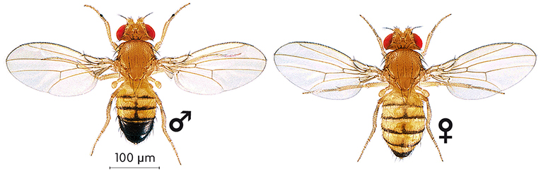
The use of the fruit fly (Drosophila melanogaster) as an experimentation organism started more than a century ago. Since then, the introduction of model animals in scientific research has been essential in order to understand the function of genes and in which way do these integrate themselves in different biological processes. Until very recently, the Drosophila has been used almost exclusively in basic research, but gradually its use has been increasing in biomedical research. More than 60% of the genes involved in human diseases, and also more than 70% of the genes related to cancer, show an equivalent in Drosophila. Moreover, the genome of this small fly can be considered a compact and simplified version of the human genome. The high levels of preservation has made possible for researchers to study the function of genes causing diseases using genetic models in flies and extrapolating these discoveries to vertebrate models and humans. On the other hand, the study of human diseases (Parkinson’s, Huntington’s or Alzheimer’s) in flies has made the study of the pathogenic routes of the disease possible and has provided new targets for the development of efficient drugs against these diseases. OUR HEALTH AND THE GENES OF A FLY The 46 human chromosomes have almost 3 billions of base pairs in the DNA organised in, approximately, 20.000-25.000 protein encoding genes. When some of these genes suffer a mutation it is usual for molecular alterations to appear and become apparent as a pathology. Many common diseases have a genetic basis, like predisposition towards diabetes, asthma, cancer or mental illnesses. Other genetic disorders are the result of the mutation of just one gene, like Huntington’s disease. In all of these cases, to the personal and familiar drama that goes with them we have to add the healthcare costs of the treatments, usually symptomatic and chronic. What can a 2-millimetre insect contribute to the development of effective treatments and possible healings? Drosophila melanogaster is a harmless insect for both humans and plants. These flies go unnoticed for most people— they feed on decomposing fruit—, but they are an essential tool for genetic research. The main advantage is that the study of any biological process in flies is simple compared to the study of vertebrates, in terms of time and costs. Besides, there is a great variety of genetic tools for Drosophila that enable us to eliminate genes, express them in tissues in which they are not usually found, mark individual cells or look for genes functionally related to a specific one, among other possibilities. To have access to experimentation organisms like Drosophila is specially important in biomedical studies, since the complete sequence of the human genome only enable us to have access to the words that constitute the genetic instructions, but not to the meaning of these instructions. In order to know how our genome works it is necessary to approach the study of the functions of each gene and their relationships with other genes and with their environment in an experimental way. Over 75% of human genes involved in diseases can be identified on flies, where their basic function is preserved. Thus, in most cases, mutations in the genes of the flies equivalent to those human genes which are responsible for human diseases (with the same origins and, generally, with the same function) produce alterations (what is known as mutant phenotype) that can be compared to the symptoms observed in patients. The availability of flies that reproduce some aspects of a human disease provides a model through which to look for new potential therapies and research the route of pathogenesis of this disease. The cycle of life of a Drosophila is of only eleven days at 25ºC, which enables us to complete experiments with relatively quickly. Thus, while the neurodegeneration characteristic of Alzheimer’s disease or Parkinson’s can take months or years to appear in models in mice, these alterations appear in a few weeks in Drosophila. |
© N. Gomple, 2008 «Many common diseases have a genetic basis, like predisposition towards diabetes, asthma, cancer or mental illnesses» |
|
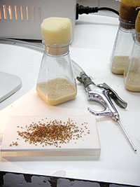
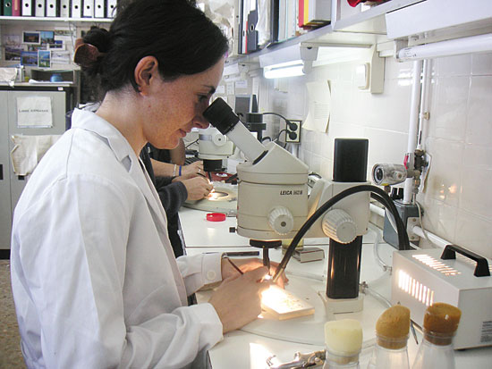 © Juan Manuel Fernández Costa The use of the fruit fly (Drosophila melanogaster) as an object of research is spread in basic research and increasingly in biomedicine. Above, a researcher from the University of Valencia observes phenotypes of Drosophila with a magnifier. Flies are anesthetised with a CO2 system, as in the picture on the right, in order to be able to observe it under the magnifier. Once flies go back to the bottles with their nurturing environment, these wake up within a few seconds. |
«El principal avantatge de Drosophila melanogaster és que l’estudi de qualsevol procés biològic es veu simplificat respecte a l’estudi en vertebrats» | |
|
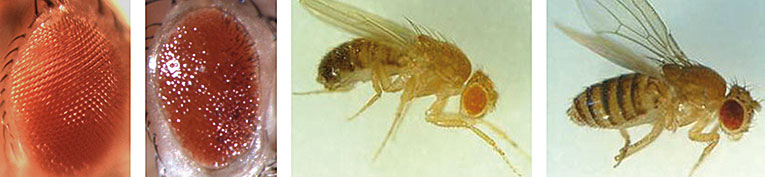
TO DIE FOR A CAUSE: NEURODEGENERATIVE DISEASES Neurodegenerative diseases are characterised by the deterioration of the performance of neurons and for being slow and gradual. Incidence in human population has alarmingly increased concurrent with the increase of life expectancy. A great deal of neurodegenerative disorders are based on mutations that affect the folding of specific proteins, which makes them accumulate in a toxic way in neurones. Biologically, the architecture of the nervous system of Drosophila is similar to that of the mammals, with areas separating specific functions like vision, learning and memory. Moreover, many of the human genes related to the neurodegenerative processes have equivalents in flies; therefore, it is not strange that these models in Drosophila were pioneers. Huntington’s disease George Huntington described in 1872 a disease that was characterised by the progressive loss of cognitive functions and changes in personality, as well as body and facial spasmodic movements. The genetic cause is on a repetitive expansion of a sequence of three nucleotides, of the CAG type, in the Huntingtine gene. The ribosomes in the cell, in charge of the synthesis of proteins using the encoded information in the RNA messengers (RNAm), translate these CAG triplets into glutamines (abbreviated to Q), one of the twenty aminoacids that build up proteins. Thus, while the huntingtine protein contains normally 8-25 consecutive glutamines, those who suffer from Huntington’s disease have huntingtine proteins with 40 or more consecutive glutamines. The existence of expansions of glutamines in at least another ten pathologies (among which there are included, moreover, different spinocerebellar ataxias, spinobulbar muscular atrophy and dentatorubropallidoluysian atrophy) has led to group them in one category of neurodegenerative disorders that share some characteristics known as polyQ diseases. The nature and common behaviour of mutations due to the expansion of CAG sequences led researchers to design a mutant variant of the Huntingtine gene and to introduce it in the genome of the Drosophila, in order to create a model in which to study the neurodegeneration typical of ployQ diseases. Flies genetically modified that expressed the mutant huntingtine protein showed a neurodegeneration, as it happens to those who suffer from Huntington’s disease, and reproduced different important aspects of the disease. First of all, the pathogenicity of the glutamine repeats depended on the length of the expansion. Thus, repeats of up to 27 glutamines caused no symptoms to humans or flies, while repeats of up to 40 glutamines caused symptoms to both of them. As in human neurodegenerative diseases, in Drosophila the degeneration was evident in the later stages of development. Often, they appeared in adults and were gradual (flies that expressed this protein lost photoreceptor neurons during their whole lives, even causing a premature death). In the same way, the presence of protein aggregates in neurons, characteristic of Huntington’s disease, also appeared in the brains of these flies. This model of Huntington’s disease developed in Drosophila has made possible to find the implication and the action mechanism of some proteins in the pathogenesis of the disease and has given new potential targets to treat it. |
 © Juan Manuel Fernández Costa «This model of Huntington’s disease developed in Drosophila has made possible to find the implication and the action mechanism of some proteins in the disease»
|
|
 |
||
|
TOXIC RNA RESPOSIBLE FOR MYOTONIC DYSTROPHY Among the diseases caused by the expansion of short sequences there is a group in which repeats are localised in non-coding regions of genes (in genes there are coding regions which determine the sequence of aminoacids in proteins, and non-coding regions with other regulating functions) as in the case of myotonic dystrophy, whose molecular bases are studied in the Group of Translational Genomics of the University of Valencia. Myotonic dystrophy is the most common kind of adult muscular dystrophy. This disease affects mainly the muscular tissue producing myotonia (difficulty in relaxing the musculature after a voluntary contraction) and muscular weakness, although being multisystemic these symptoms appear usually together with others, like iridescent cataracts, cardiac arrhythmia or gastrointestinal disorders. The mutation responsible for the myotonic dystrophy is placed in a non-coding region of the gen DMPK (Dystrophia Myotonica Protein Kinase) and is made up of repeats of the sequence CTG. In normal conditions, of these triplets we find between 5 and 37 copies, while in patients with myotonic dystrophy the number of copies increases, even up to 3000. Different animal models of the disease, including a model in Drosophila created by our group, has made possible to show that myotonic dystrophy originates because the RNAm with a great number of CTG sequences become toxic for the cell, regardless of what is the protein they are encoding, which constitutes a new mechanism of pathogenesis. Why are these RNAm toxic? It is known that different proteins which are critical for the metabolism of nucleic acids join to these RNAm in an abnormal way and are sequestered leaving them unable to perform their normal functions in the cell nucleus. Among them, the sequestration of a series of proteins named muscleblind is critical for the development of symptoms in the disease. What happens if we express human mutations in flies? In our group we have created transgenic flies that express 480 repeats of the CTG sequence in different tissues of Drosophila. Surprisingly, although a mutation of this kind has never been described in this insect, the expression of a compound RNAm produced exclusively by CTG repeats reproduces typical molecular, histological and degenerative aspects of the human disease. Not only is this mutation capable of altering the operation of the cells of the insect, but also a copy of the human gene muscleblind is capable of operating in Drosophila. Both experiments are an example of what we can call the «humanisation» of the genome of Drosophila, which is contributing to a better understanding of some human pathologies and which can help to find therapies for patients affected by these. DISCOVERY OF DRUGS IN DROSOPHILA Nowadays, the development of a medicine and its marketing costs more than 800 million dollars and it takes an average of fifteen years to go to market. A great deal of these costs is caused by the lack of effectiveness or the toxicity of the candidate compounds in the preclinical and clinical stages of their development. This is the reason why there is a great interest in identifying compounds with a therapeutic potential for the palliative treatment of genetic diseases in preclinical stages. To carry out a massive in vivo research of compounds in a simple invertebrate organism like Drosophila enables us not only to identify molecules that improve one aspect of the pathology, but also to classify them according to their pharmacological characteristics. A compound, aside from having a specific biological activity over a cell process altered by a pathology, has to have optimum pharmacological properties, with a good absorption, distribution or low toxicity, among others. Many in vitro or cell culture tests do not take into account these pharmacological characteristics due to the inherent limitations of the systems used. In this respect, drug research in Drosophila is twice as demanding. On the one hand, the identified compounds have to improve the symptoms that reproduce the human disease. On the other hand, the use of this compound has to be compatible with the life of an organism that, genetically, is a simplified version of he human genome. In most cases, biologically active compounds identified in Drosophila operate in their environment in a similar way in mammal cells. A spin-off of our research group, Valencia Biopharma, has accepted the challenge of discovering potentially therapeutic drugs using this strategy. Begoña Cabia Fernández. Pre-doctorate scholarship holder. Laboratory of Molecular and Cellular Endocrinology. Research Institute of Health. University Hospital Complex of Santiago de Compostela (CHUS). |
© Mètode «Biologically, the architecture of the nervous system of Drosophila is similar to that of the mammals, with areas separating specific functions like vision, learning and memory» |
|
BIBLIOGRAPHY
Bilen, J.and N. M. Bonini, 2005. «Drosophila as a Model for Human Neurodegenerative Diseases». Annual Review of Genetics, 39: 153-171.
García López, A. et al., 2008. «Genetic and Chemical Modifiers of a CUG Toxicity Model in Drosophila». PLoS One, 3(2): e1595.
Marsh, J. L.andL. M. Thompson, 2004. «Can Flies Help Humans Treat Neurodegenerative Diseases?». Bioessays, 26(5): 485-96.
Reiter, L. T. et al., 2001. «A Systematic Analysis of Human Disease-Associated Gene Sequences in Drosophila melanogaster». Genome Research, 11(6): 1114-1125.