Rudolf Nieuwenhuys
«Science is tremendously susceptible to fashion»
Emeritus Professor of Neuroanatomy at the Netherlands Institute for Neuroscience
Rudolf Nieuwenhuys is a worldwide eminence in the field of Neuroanatomy. He has a formal education as a medical doctor, but his view is not at all anthropocentric and he has always been fascinated by the great diversity of forms found in the brain of different animals. He was a pioneer in using embryonic development as a fundamental principle in order to understand brain structure and evolution. He considers himself privileged for being able to work at his age, 88 years old. His insatiable curiosity has not been diminished with age and his mind remains open to new ideas, even if these imply reconsidering old settled dogmas. He is not afraid of new challenges and gets excited in front of each new finding.
«I realized that all these brains shared a common basic structural plan. That was the beginning of my interest in comparative neuroanatomy»
It is Saturday morning, and Professor Nieuwenhuys made an appointment with us at 10 am at his house in Abcoude, a small town near Amsterdam. It is cold and rainy, a typical late January day in these latitudes. We walk along the canals following a map skillfully drawn by Suzanne Baker (his partner) the previous evening, when both of them picked us up at the airport. We arrive on time to the house located in a quiet, residential area. Suzanne opens the door and guides us to a huge and luminous room, including living room, library, dinning room, and kitchen. In spite of its large dimensions, the room is very cozy and stylish. There are several aerial photographs of different cathedrals on the walls. Later we will learn about the professor’s fascination for these temples and his trips around different European cities in order to make photographs of them from different angles, including from above thanks to Suzanne, since she is a wonderful aircraft pilot (now we understand the highly detailed and accurate map she sketched the previous evening). Before the interview, and after checking that Suzanne is not listening, the professor proudly explains us how extraordinary she is, how much they complement each other intellectually, and how lucky he is for being able to count on her moral and practical support to carry out his projects.
The professor receives us and we get around an enormous table covered in books and papers that he will use during the interview to illustrate his explanations. He has minutely prepared the interview, with the high level of detail and perfection that fills all his work. During more than two hours he will talk about his life, which is part of neuroscience history, and about his present and future projects.
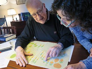
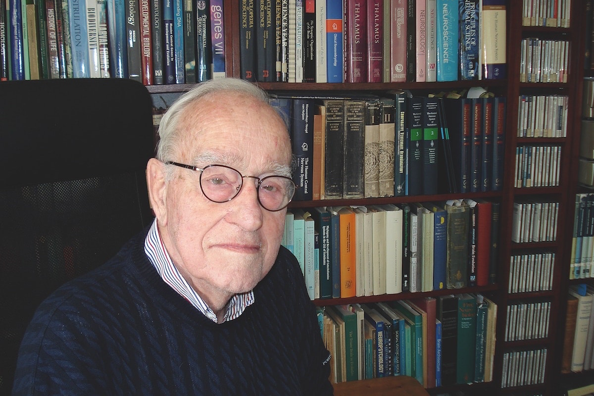
Photo: Ester Desfilis
How did you become interested in research? And why did you start to do comparative neuroanatomy studies?
I studied Medicine during the years just after the Second World War. During the war and the German occupation, many universities in Holland were closed and there was an enormous post-war affluence of medical and other students to the universities. Because of the limited capacity in the various clinics, there was a long waiting time of about a year between the end of the theoretical training and the beginning of the practical clinical curriculum. Then, I decided to take a look at the Netherlands Central Institute for Brain Research [in Amsterdam]. I was passing in front of that institute every day when going to the anatomy department, but the door was always closed and quite mysterious. A certain day I said «well, I will ring the bell» and there were only two persons working there, a technician and an administrator. The director of the institute, the world famous Professor C. U. Ariëns Kappers, had died six years before, in 1946. After his death the institute was just dormant. The two «survivors» were extremely kind with me and showed me the institute, including the immense collection of brains preserved as a whole and in sections, and the library of the professor. I was deeply impressed. These two people spoke with the highest respect about the professor and all that he had done. As a medical student, I was at that time somewhat familiar with the human brain, but then I had the opportunity to look at brains of all sorts of vertebrates. I gradually realized that all these brains, however different in size and shape, shared a common basic structural plan. That was the beginning of my interest in comparative neuroanatomy.
In any case, it is not common to find a medical doctor with such an ample interest and knowledge about brain evolution. What were your first steps in comparative brain research?
When I finished my medical studies, I became employed at the institute as a young assistant, and started to work on a doctoral thesis. A physiologist called Jaap Segaar was working on the neural substrate of the reproductive behavior of the three-spined stickleback, a small bony fish. The male of this species shows a very interesting reproductive behavior, building and guarding a nest where the female spawns her eggs that he fertilizes. Segaar found that when you removed the entire telencephalon, the fish started to build a nest by digging a hole but did not know how to proceed. Being a physiologist he had no idea of the microscopic structure of the brain, and he asked me for help. That became the subject of my PhD thesis. I found that the development of the forebrain of the stickleback, and that of all bony fishes, differs considerably from all other vertebrates, and this difference strongly influences the interpretation of forebrain organization. I was trained as a medical doctor and I took, in addition, many courses in zoology. That position, on one hand being a medical doctor and on the other hand being a biologist, has remained with me throughout my career; sometimes I focus entirely on a biological problem and other times I am on the medical side. That is why I call myself a «bicomponential» (an entirely new word!).
«That position, on one hand being a medical doctor and on the other hand being a biologist, has remained with me throughout my career»
What would you say to a young fellow interested in comparative studies?
When I gradually became a comparative neuroanatomist, many people said to me (intending to really give me good advice): «But Rudolf, what are you doing now? You have a wife and two small children and you have to think about your future, and this would be absolutely foolish. There is no future whatsoever in comparative neuroanatomy, because it is all known and all finished». Three years later, I went for one year (1961-1962) as a postdoc to the United States. Having done just the thesis, I was at the annual meeting of the American Anatomical Society and seven heads of department offered me a job! But I decided to go back to the Netherlands. At that time, comparative studies were a boom in the USA. Science is tremendously susceptible to fashion. At a certain time, a given discipline is in, and later it is out and nobody can ascertain why that is. So, it is very hard to advice young people to do this or that. My advice is: do what you would like to do, and hope for the best.
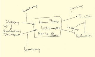
Figure 1. Diagram drawn by the professor during the interview, explaining the inter-relation between ontogeny/development, evolution, function and dysfunction, and how all of these contribute to the «understanding» of the human brain.
You have contributed a relevant textbook on human neuroanatomy1 that has become a reference in many medical schools around the world (it has even been translated to Chinese and Japanese). The work differs from others because of the inclusion of abundant data from brain research in different species, including non-mammals. Why do you think comparative studies are important for understanding human brain organization?
Teaching human neuroanatomy or writing a book about that subject for the medical profession is always difficult. The human brain is so ultra-complex that high quality figures, both diagrammatic and realistic, are indispensable (hence, the inclusion of the artist as co-author). The human brain is the product of two highly inter-related processes, its ontogeny and its evolutionary development. Knowledge of these processes and their interdependence is conditio sine qua non for the understanding of the human brain, but the time (in lectures on the human nervous system) or the space (in books on the same subject) for these fundamental subjects is limited. I taught human neuroanatomy for 25 years and, until the end of my career I struggled with the problem of how to explain the brain, so that they are not just learning by heart. You can know this is here and this is there, but «understanding» is extremely difficult. The pertinent lectures or books should also cover the functional significance of the structures and the clinical implications of the morphological facts presented. Thus, here you see the real dilemma, to tell how it is, to give the functional and the clinical implications as much as possible, and in addition to give an idea about its ontogenesis and evolution. [While he answers the question, he makes a drawing explaining these ideas that is reproduced in Figure 1.]
«The human brain is the product of two highly inter-related processes, ontogenyAnd evolutionary development. Knowledge of these processes is ‘conditio sine qua non’ for the understanding of the human brain»
The department you chaired during more than 20 years2 became a worldwide reference for comparative neuroanatomy studies. However, this tradition started to fade off at the turn of the century. How do you explain the decline of this tradition in your former department?
I was attracted by the Department of Anatomy of the Faculty of Medical Sciences of the University of Nijmegen not because I was a comparative neuroanatomist, but simply because they needed a person who was able to manage the research of that department. At that time, I had done a substantial amount of comparative studies and that’s not too surprising because I was working at a brain institute only devoted to research, and I had 95% of my time for research. Thus, I went to the department and the comparative school of Nijmegen gradually got some fame. I had more than thirty different graduate students doing a doctoral thesis under my supervision. But I hated to go to faculty meetings, and my senior technician, who was much better in that than me, said to me at the end of my career: «You are a very good scientist but you are a poor politician and you will see, now they do not dare to do that, but after your retirement they will strongly attack and reduce the department of anatomy». And exactly that happened. At the time of my farewell, there were 54 people working in the lab, and within a few years it was reduced to twelve, and that was unfortunately the end of the comparative school in Nijmegen.
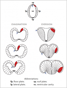
Figure 2. Scheme of the developing forebrain (in particular, the telencephalon) from the neural tube employed here to explain the topology concept. During morphogenesis, the telencephalon may grow following two different processes: evagination (this happens in most vertebrates) or eversion (only in teleost fishes). As a consequence, the same brain subdivision (as that highlighted in red) can finally occupy different topographic positions (either close to or far from the midline), but the topological position is identical (adjacent to the roof plate; pointed with an arrow).
You were the first in introducing the concept of topology (from the field of mathematics) for studying and comparing the brain of different vertebrates. This concept has become fundamental in studies of brain development and evolution. How would you explain this concept to the general public?
That is not easy. It has to do with transformations. Topology is a branch of mathematics that studies those features in figures that remain unchanged when you transform things in all sorts of directions. I have applied only the elementary principles of topology into neuromorphology. I can explain that with an example, the development of the forebrain, which is shown in this figure [the professor continues the explanation referring to Figure 2]. During early development, this and all other parts of the brain, have the shape of a simple tube. A cross section of this tube is shown in the figure. This tube has thin side walls, named lateral plates, which on the upper side are interconnected by a narrow strip, the roof plate. The lines of attachment between the lateral plates and the roof plates are known as the taeniae [indicated by arrows in the figure]. During further development, two entirely different morphogenetic processes may manifest: evagination and eversion. During evagination, the lateral plates bulge out and are going to surround ventricular cavities. The roof plate remains narrow. The adult forebrains of almost all groups of vertebrates are the product of this evagination process. Eversion, on the other hand, is confined to bony fishes. During this process the lateral plates fold to the sides with consequent widening of the roof plate. Ultimately, this structure becomes a thin membrane, which surrounds a large portion of the forebrain. We say that the brain areas situated directly adjacent to the taeniae (giving rise to the hippocampus in many vertebrates, shown in red in the figure), occupy in adult evaginated and everted brains entirely different topographical but corresponding topological positions. Because the shape of the wall of the brain shows many variations, the general rule is that structures in the brains of different animals should not be compared on the basis of their topographical, but rather their topological, positions. Neglect of this rule has led to much confusion in comparative neuroanatomy.
In 1998, you published, together with ten Donkelaar and Nicholson, a three-volume handbook entitled The central nervous system of vertebrates. What made you undertake this huge project? What do you think is the main contribution of this opus magnum?
At the time when I started out as a comparative neuroanatomist, the only comprehensive reference book was the work of Ariëns Kappers, Huber and Crosby, which appeared in 1936. This work was not only outdated; it also had some serious shortcomings. The plan for a new and better reference work arose gradually during my professorship at the University of Nijmegen. I wrote an outline for the book, which all collaborators agreed with. We agreed that: the book should rest on a topological basis; the importance of the study of the development for the understanding of the structure of the adult brains should be emphasized; the book should be coherent, that is to say: all chapters on the brains of the various groups of vertebrates should follow the same pattern, with regard to both text and illustrations; and of course, an enormous amount of new data should be included. During the second half of the twentieth century, quite a great number of techniques made it possible to study the fiber connections, and so everybody was doing experimental studies on fiber tracts in all sorts of vertebrates. Here you can see how much techniques dominate a whole discipline. In the book we covered all that. It is also important to note that in those days the medical faculty in Nijmegen included a department of illustration, where ten highly competent artists were working, and I took advantage of it.
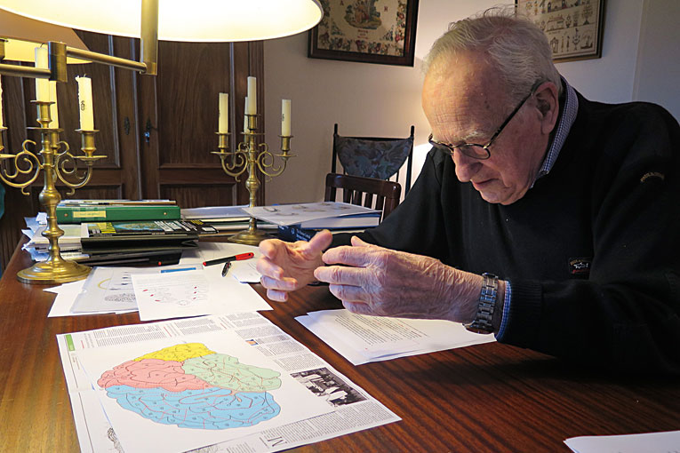
Photo: Ester Desfilis
With the boom of molecular genetics and the use of the expression patterns of developmental regulatory genes for studying the brain, some researchers are using gene expression as support for homologies (sometimes as the only criterion). What do you think should be the role of topology in the interpretation of these data?
The topological relations are still of paramount importance for establishing homologies; however, the modern molecular approach can help us to disclose relationships that we could not see previously. When I wrote the book in 1998, the molecular approach was just beginning. Nobody knew at that time that there would be an enormous development of molecular developmental neuromorphology. But it happened that 15 years later I was invited to give a series of lectures on this subject in Leipzig (Germany). Following these lectures a book was developed, which was just published. The book is entitled Towards a new neuromorphology, in collaboration with the noted Spanish neuroscientist Luis Puelles (University of Murcia). In that book we present a survey of these new developments which have an impact on neuromorphology. Our conclusion is that the study of the localization of developmental regulatory genes and their products (genoarchitecture) is extremely helpful in the recognition of hitherto obscure topological relations.
«Modern molecular developmental neurobiology has shown many deep and real correspondences that could not be detected with classical techniques»
Comparing the expression and function of these genes between vertebrates and invertebrates has led some authors to propose the concept of deep homology, for instance for comparing the eyes of vertebrates and invertebrates. What do you think about this concept?
First of all, I would like to say that I am not a specialist in genomics. So, my answer can only be limited. I believe that modern molecular developmental neurobiology has shown many deep and real correspondences that could not be detected with classical techniques, and I do believe that these deep homologies exist and I do believe that this opens the way for comparisons between vertebrates and invertebrates in a way that was formerly absolutely impossible. Based on molecular data, some researchers now make comparisons between the high cognitive centers in the brain of insects, called the mushroom bodies, and the cerebral cortex. This is intriguing, but I am simply not in a position to judge whether these comparisons are correct or not.
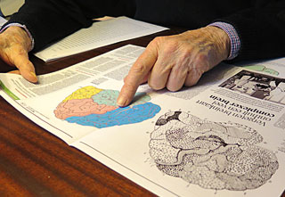
Figure 3. Professor Nieuwenhuys compares the Brodmann map with the new one he recently published, which is based on myeloarchitecture and may thus be useful for interpreting neuroimaging data. / Photo: Ester Desfilis
What other projects are you currently working on?
Things in science are never completely determined for a long term. You encounter, sometimes quite unexpectedly, new problems, and that happened to me a couple of years ago when I became involved in a new branch of neuroscience, neuroimaging. I said before that the directions of science are to a very large extent dictated by new techniques, more than by new concepts, and we could say grosso modo that we have now two new lines, the molecular one explained above, and the imaging techniques. At the time that these techniques first appeared there were headings in newspapers and even in scientific journals saying: «We can now look into the living brain and we can even see and study the mind.» I was also impressed, but I also saw right from the beginning that when you are asking a person to do something, for instance, think a while about your mother in law, or make an equation, and you get activity in the brain, but this is not yet science. It becomes science, and only the beginning of science, if you can tell where that particular activity is situated. To localize the activity, they took the best known map of the cerebral cortex, the one by Brodmann3. This is still the main approach in finding the relation between cortical function and cortical structure. The new development of MRI (magnetic resonance imaging) allows to obtain functional and structural information about the same brain, which is a tremendous leap forward. However, by looking at the images you can see absolutely nothing of the arrangements of cell bodies (cytoarchitecture), the feature on which Brodmann’s map is based. But you can see at high resolution the arrangement of the myelinated fibers in the cortex very well, what is called the myeloarchitecture. Then, I realized that a cerebral map based on myeloarchitecture was needed. That made me revise the work of the German school of Vogt and Vogt, in which Brodmann was working when he produced his famous map. My conclusion was that the work was absolutely sound and that these people had accumulated enough material to make a map of the human cortex. Then I formed a team with a person who was very good in MRI and another person who was very good in computer programming. Finally, we got a standard map of the myeloarchitecture of the human cortex, in which 180 fields can be recognized. This map has been published this year [the professor shows us the new map in Figure 3], and now we are making other maps of other features. In addition, I am part of an international group of six persons, called «bridging the gap». This group is coming together four times a year, sitting around this very table, to discuss the problem of the relation between the structure and function of the cerebral cortex.
At the end, we have the feeling of having been with a highly educated person, fascinated by knowledge, and with an extraordinary capacity for relating ideas and «bridging gaps» to generate novel perspectives for understanding human brain.
1. Nieuwenhuys, R., Voogd, J., & van Huijzen, Ch. (2009). The human central nervous system, 4th ed. New York: Springer (Go back)
2. In 1968, he moved to the University of Nijmegen (the Netherlands), where he held the Chair of Human Neuroanatomy in the Faculty of Medical Sciences, and the Chair of Comparative Neuroanatomy in the Faculty of Natural SEl 1968, es va incorporar a la Universitat de Nimega (Països Baixos), on va ocupar la Càtedra de Neuroanatomia Humana a la Facultat de Medicina i la de Neuroanatomia Comparada a la Facultat de Ciències Naturals. Es va retirar d’ambdues càtedres el 1992. (Go back)
3. Published in 1909. (Go back)